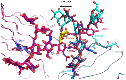
Fig. 4.
Overlay of view on the interaction interface between glycosylated Fc receptor and Fc fragment. Chain A of the afucosylated Fc fragment is shown in blue, its complexed Fc receptor in cyan. Chain A of the fucosylated Fc fragment is shown in magenta, with core fucose highlighted in yellow; its complexed Fc receptor is in dark violet.

