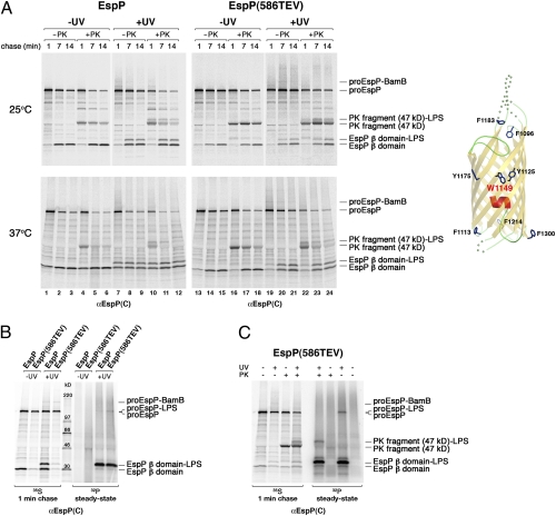
Fig. 4.
Residue 1149 is exposed to lipid after the initiation of passenger domain secretion. (A) AD202 cells were transformed with pDULEBpa and a derivative of pRI22 or pRI23 harboring an amber codon at residue 1149. Cells were subjected to pulse-chase labeling at 25 °C or 37 °C after the addition of 200 μM IPTG, and half of each sample was UV irradiated. Both UV-irradiated and nonirradiated samples were divided in half, and one half was treated with PK. Equal portions of all the samples then were used for immunoprecipitations with an anti-EspP C-terminal antiserum. The crystal structure of the β domain and the location of residue 1149 are shown in the diagram on the right. (B and C) The experiment in A was repeated, except that a culture containing KH232PO4 was grown in parallel. UV irradiation (B) or both UV irradiation and PK treatment (C) of the 35S- and 32P-labeled cells was conducted as described above. Equal portions of all the samples were used for immunoprecipitations with an anti-EspP C-terminal antiserum.

