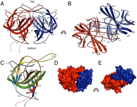
Fig. 1.
Crystal structure of astrovirus P2415–646. (A) Side view of the P2415–646 dimer. The two subunits are colored in red and blue, respectively. The N and C termini of the red subunit are highlighted by dotted lines. The viral capsid lies at the bottom the dimeric spike. (B) Top view of the P2415–646 dimer. (C) Structure of one P2415–646 molecule. The polypeptide is rainbow-colored with the N terminus in blue and the C terminus in red. (D and E) Surface representations of the P2415–646 dimer for comparison with the cryo-EM reconstruction image. Molecules in D and E are viewed from the same orientation as in A and B, respectively. The figures were made with PyMOL (39).

