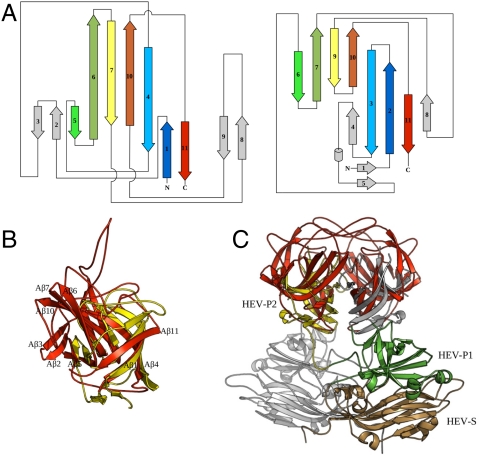
Fig. 2.
Structural comparison of P2415–646 with HEV capsid P2 domain. (A) Topology diagrams for the astrovirus P2415–646 (Left) and the HEV P2 domain (Right). Related β-strands in the two structures are rendered in matching colors, and unrelated structural elements are shaded in gray. (B) Superposition of the astrovirus P2415–646 (red) and the HEV P2 domain (yellow). Structure elements in astrovirus P2415–646 are labeled as in Fig. 1C with a letter “A.” (C) Superposition of the atrovirus P2415–646 dimer with the HEV CP dimer. The atrovirus P2415–646 dimer is in red. For HEV CP, one subunit is in gray, and the other is colored by domains: S in gold, P1 in green, and P2 in yellow.

