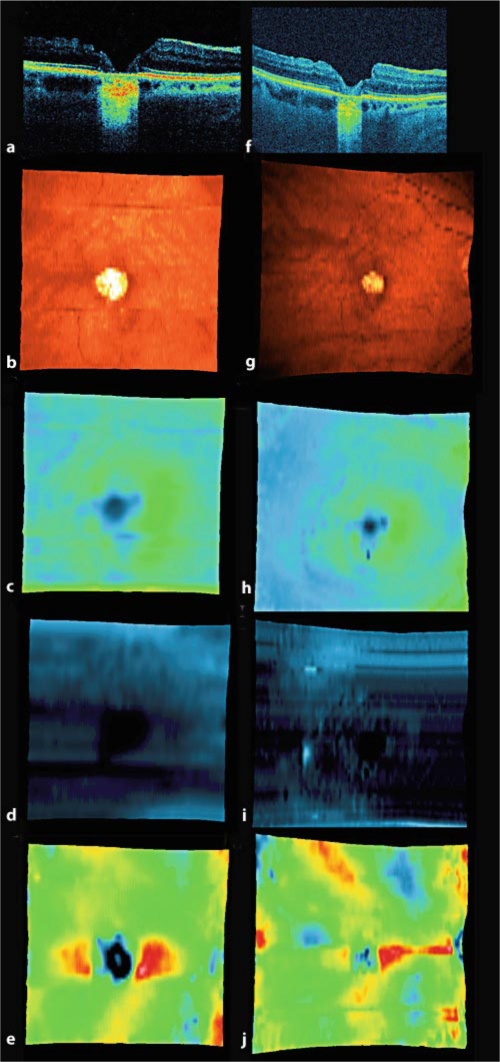
Fig. 2.
Patient 2. SD-OCT and SD-OCT fundus maps 1 week (a-e) and 3 years (f-j) after macular hole surgery to join the hole borders. Decrease in size of the RPE defect may be observed. a, f SD-OCT B-scan performed at the same level of the retina. b, g SD-OCT fundus map. c, h SD-OCT retinal thickness map, darker colors mean thinner retinal tissue. d, i SD-OCT nerve fiber layer map, darker colors mean thinner retinal tissue. e, j SD-OCT RPE map, darker colors mean thinner retinal tissue.

