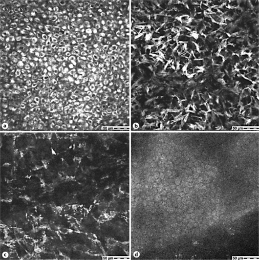
Fig. 2.
Confocal microscopy of the cornea (right eye) shows highly reflective intracellular deposits in the epithelium (a): depth 5 μm, anterior stromal keratocytes (b): depth 52 μm and mid stromal keratocytes (c): depth 354 μm. d The endothelium appears regular in morphology and without significant intracellular deposits.

