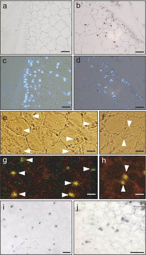
FIGURE 1.
Plant cells accumulate iron in the nucleus. a–j, histochemical analysis of pea embryonic cells (a–d), A. thaliana (i), and tomato leaf mesophyll cells (j). e–h, elemental imaging was performed on sections of pea embryos embedded in resin. a, negative control of the Perls/DAB staining where the first reaction with potassium ferrocyanide was omitted. b, Perls/DAB staining of iron. c, DAPI-stained nuclei revealed by epifluorescence (from control section shown in panel a). d, merge of Perls/DAB and DAPI reactions. e and f, bright field microscopy of unstained cells. g and h, μPIXE analysis of the same sections with iron imaging in green and phosphorus imaging in red. Samples were scanned with a 3.0-MeV proton beam focused to 1-μm size and measuring the X-rays emitted at 6.4 keV for iron and at 2.0 KeV for phosphorus. White arrows point out the cell nucleus. Bar = 40 μm for panels a–d and i–j, bar = 20 μm for panels e–h.

