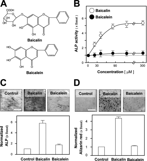
FIGURE 2.
Stimulatory effect of baicalin and baicalein on osteogenesis in cultured osteoblasts. A, chemical structure of baicalin and its aglycone baicalein. B, application of baicalin in cultured osteoblasts for 3 days increased ALP activity in a dose-dependent manner. Application of baicalein for the same period of time did not show induction effect on ALP activity. C, application of baicalin (50 μm) and baicalein (50 μm) for 3 days. The ALP amount was quantified by histochemical staining. D, cultured osteoblasts were able to undergo mineralization upon application of baicalin (50 μm) in the presence of β-glycerophosphate (5 mm). After 21 days of the treatment, the stained nodules were found, as shown by Alizarin Red staining (upper panel). The degree of mineralization after treatment of baicalein (50 μm) was not obvious. Alizarin Red was quantified using a solution of 20% methanol and 10% acetic acid in water and read on a spectrophotometer at 450 nm (lower panel). Values in all panels are expressed as the fold of increase to basal reading (control culture; 0.02% DMSO) and are means ± S.E., n = 5, each with triplicate samples. Scale bar, 5 mm.

