FIGURE 8.
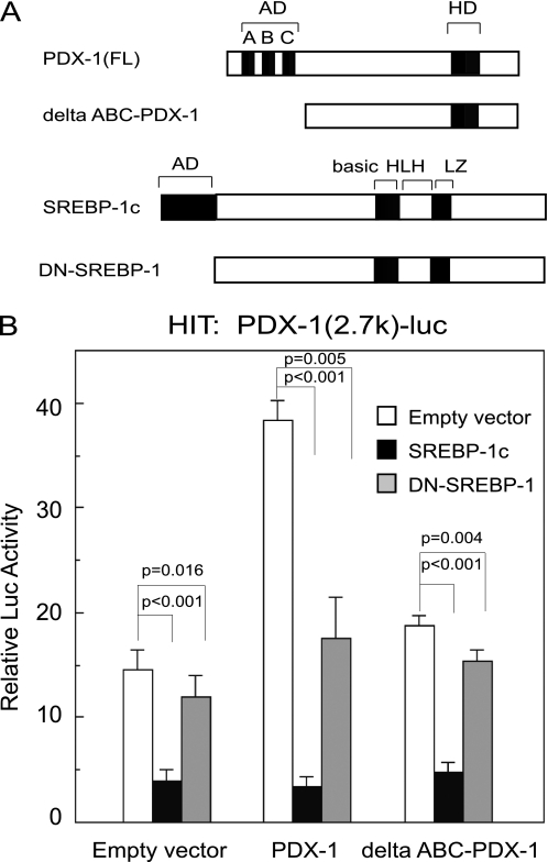
Physical interaction between PDX-1 and SREBP-1c partially contributes to SREBP-1c suppression of the Pdx-1 promoter. A, schematic representation of PDX-1, Δ-ABC-PDX-1, SREBP-1c, and DN-SREBP-1c. Δ-ABC-PDX-1 lacks 75 amino acids in the N-terminal transactivation domain of PDX-1. DN-(Δ-1–67)-SREBP-1c lacks 67 amino acids in N-terminal transactivation domain of SREBP-1c. B, PDX-1 (−2.7 k)-Luc (0.5 μg) or the empty vector pGL3-Luc (0.5 μg) and pSV-β-gal (0.5 μg) were cotransfected with CMV-PDX-1 (0.25 μg), CMV-ΔABC-PDX-1 (0.25 μg), CMV-SREBP-1c (0.25 μg), and CMV-DN-SREBP-1 (0.25 μg) in HIT cells (n = 6). Luciferase activity was normalized to the pSV-β-gal values. The pGL3-Luc activity of empty expression vector (CMV-7) was set to 1.0. Data are means ± S.E. Statistical significance was assessed using the Student's t test for unpaired data.

