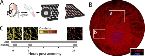
FIGURE 1.
In vitro axotomy model. A, DRGs were dissected from E12.5 mouse embryos and dissociated in trypsin (5 × 10⁵ neurons/ml). Cell suspensions were delivered as single 0.5-μl droplets to the dry laminin/PDL-coated surface of each well in a 96-well microtiter plate with a liquid handling machine. Medium was then added after cells had adhered to a 1–2-mm portion of the well. B, montage of DRG spot culture at 7 days in vitro. Boxes indicate imaging regions (a, distal/injured; b, proximal/uninjured); red = β-tubulin (Tuj1 antibody), blue = DAPI. Dashed white line indicates where axons are cut (axotomy). Well diameter = 7 mm. C, screening time line: after a 30-min preincubation with compound, axons were severed with a blade, and axon integrity was quantified from brightfield images of axons taken at 0, 6, and 24 h post-axotomy. Axon fragmentation was quantified from each image (see supplemental Fig. S1). Representative images are pseudo-colored by particle circularity (see “Experimental Procedures”).

