FIGURE 3.
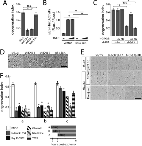
IKK and GSK3 are required for rapid axon degeneration. A, knockdown of murine IKKβ by shRNA (shIKKβ 1, 2) suppressed axon degeneration measured at 9 h post-axotomy. Expression of dominant-negative IκBα had no effect. B, dominant-negative IκBα suppresses basal and TNFα-induced NFκB activity in HEK 293T cells. C, knockdown of murine GSK3α and GSK3β (shGsk3) by shRNA suppresses axon degeneration at 24 h post-axotomy, whereas expression of constitutively active (CA), but not kinase-dead (KD) human GSK3β (h-GSK3β) restored normal axon degeneration in this setting. D, representative images from panel A. E, representative images from panel C. F, protective effects of the indicated compounds when added at different times post-axotomy. Addition of compounds (indirubin 3′-monoxime, 5 μm; Bay 11-7082, 4 μm; gliotoxin, 2 μm; nifedipine, 10 μm; TPCK, 28 μm) 1 h prior (a) or minutes after axotomy (b) suppressed axon degeneration measured 9 h post-axotomy (each compound compared with DMSO added at the same time), whereas neither compound suppressed axon degeneration when added 2 h post-axotomy (c). Error bars show S.E.; *, p < 0.001 by Student's t test; N.S., not significant (p > 0.01); 4 replicates per group.

