FIGURE 3.
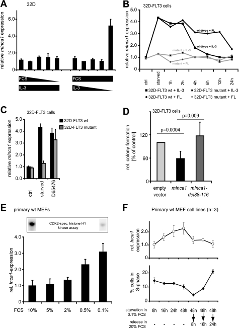
Inca1 expression is induced by serum starvation and repressed by pro-proliferative signaling. A, regulation of Inca1 mRNA expression was analyzed in IL-3-dependent 32D cells by quantitative RT-PCR. Serum starvation alone in the presence of IL-3 did not induce Inca1 mRNA expression, whereas depletion of IL-3 in the presence of serum resulted in increased Inca1 mRNA expression, indicating the dependence of Inca1 induction on the inhibition of mitogenic signaling rather than serum concentration. B, mitogenic signaling from a constitutively active mutant FLT3 receptor FLT3-ITD (36) blocked Inca1 induction as determined by quantitative RT-PCR. Stable expression of the FLT3 wild type tyrosine kinase receptor in 32D cells did not alter Inca1 induction after IL-3 depletion. Inca1 mRNA induction after IL-3 depletion was reversible either by addition of IL-3 or by addition of FLT3 ligand (FL). Expression of leukemogenic, constitutively active FLT3-internal tandem duplications (FLT3-mutant) inhibited Inca1 induction. Addition of IL-3 or FL after IL-3 starvation did not change expression of Inca1 in 32D-FLT3 mutant. C, tyrosine kinase inhibitor D65476 increased Inca1 expression in 32D-FLT3-ITD to a level similar to that detected in 32D-FLT3 after starvation. Thus, Inca1 suppression through FLT3 mutant was reversible upon tyrosine kinase inhibition, and Inca1 was induced upon inhibition of mitogenic signaling. D, inhibition of colony formation in hematopoietic progenitor cells 32D-FLT3 requires the cyclin-binding site. Colony formation assays were performed to analyze the growth inhibitory functions of murine Inca1-full length and the cyclin binding-deficient mutant del88–116. The homologous sites required for cyclin binding were mapped and deleted in murine Inca1 (see supplemental Fig. S1). Expression of murine Inca1-full length (mInca1) led to significantly decreased colony formation (p = 0.0004). No growth inhibitory effect was observed for the cyclin binding-deficient mutant mInca1-del88–116. Indicated are means ± S.D. of three independent experiments. E, serum starvation of primary MEFs dose-dependently induced expression of Inca1 mRNA. MEFs were starved for 48 h in 0.1% FCS and released for 24 h in the indicated FCS concentrations. Lower panel, expression levels were determined by quantitative RT-PCR and normalized to GAPDH expression. Upper panel, lysates from cells grown at 0.1 or 10% FCS were subjected to CDK2-specific histone H1 kinase assays and revealed high CDK2 kinase activity in cells grown with 10% FCS. F, time course of the regulation of Inca1 expression and cell cycle distribution during starvation and release of primary wild type MEFs (n = 3). The regulation of Inca1 expression preceded the re-entry of cells into S-phase after refeeding of the starved cells. The Inca1 expression was determined as in E, and cell cycle distribution was assessed by FACS analysis after BrdU incorporation and propidium iodide staining. Please note that the mean ± S.D. was derived from three independent batches of MEF cells.

