FIGURE 7.
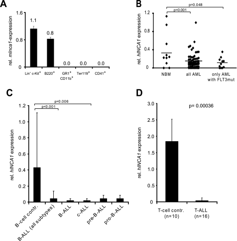
Inca1 expression is suppressed in human AML and ALL blasts. A, Inca1 mRNA expression was determined by real time quantitative RT-PCR using cDNA from murine bone marrow cells, which were sorted by FACS. GAPDH served as housekeeping gene for normalization. Inca1 was expressed in hematopoietic stem/progenitor cells (Lin− c-Kit+) and lymphoid progenitor cells (B220+). Granulocytic (CD11b+/GR1+), erythrocytic (Ter119+), or megakaryocytic progenitors (CD41+) did not express Inca1. B, INCA1 expression is significantly down-regulated in bone marrow cells from human AML patients compared with normal bone marrow cells (NBM). In bone marrow cells from AML patients, which were positively tested for the FLT3-ITD mutation, INCA1 expression was specifically and significantly less expressed compared with normal bone marrow samples. INCA1 expression was determined by quantitative RT-PCR and normalized to GAPDH expression level. C, expression of INCA1 was also significantly repressed in B-ALLs, especially in the subtype c-ALL, compared with normal B-cells. B-cell control, n = 12; ALL (all subtypes), n = 46; B-ALL, n = 10; c-ALL, n = 25; pre-B-ALL, n = 7; pro-B-ALL, n = 4. D, T-ALL bone marrow cells expressed significantly less INCA1 than normal T-cells (p = 0.00036).

