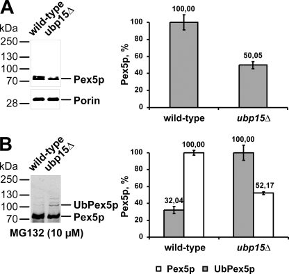
FIGURE 7.
ubp15Δ cells exhibit lower steady state concentration of Pex5p but higher rate of ubiquitinated Pex5p. A, whole cell lysates of oleic acid-induced wild-type and ubp15Δ cells were prepared and subjected to immunoblot analysis with antibodies specific for Pex5p and mitochondrial porin, which served as a loading control (left). Signal intensity was estimated by densitometric analysis (right). B, indicated strains were grown for 10 h under oleic acid conditions and for an additional 4 h under the same conditions in the presence of MG132 to inhibit proteasomal degradation. Whole cell lysates were prepared, and equal portions were subjected to immunoblot analyses with Pex5p antibodies (left). Signal intensity of modified Pex5p in ubp15pΔ cells and unmodified Pex5p in wild-type cells was quantified by densitometry (right).

