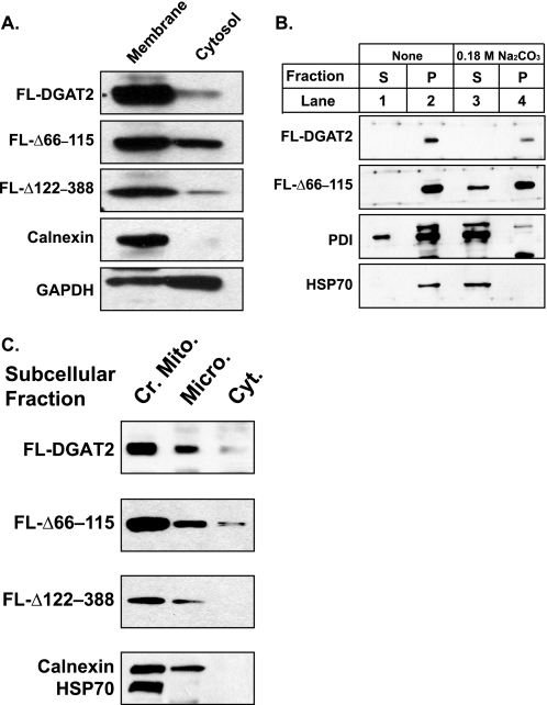
FIGURE 2.
DGAT2 associates with cellular membranes even in the absence of its transmembrane domains. A, total cellular membranes and cytosol were isolated by ultracentrifugation from HEK293T cells expressing FL-DGAT2 and the indicated DGAT2 mutants. Fractions were separated by SDS-PAGE and immunoblotted with anti-FLAG, anti-calnexin, and anti-GAPDH antibodies. B, total membranes from HEK293T cells transfected with FL-DGAT2 and FL-Δ66–115 were resuspended in PBS (lanes 1 and 2) or PBS containing 0.18 m sodium carbonate (pH 12) (lanes 3 and 4). After incubation, the pellet (P) and supernatant (S) fractions were isolated by centrifugation and analyzed by immunoblotting with anti-FLAG, anti-protein disulfide isomerase (PDI), and anti-HSP70 antibodies. C, crude mitochondrial (Cr. Mito.), microsomal (Micro.), and cytosolic (Cyt.) fractions were isolated from HEK293T cells expressing FL-DGAT2, FL-Δ122–388, or FL-Δ66–115. An equal amount of protein (15 μg) from each fraction was immunoblotted with anti-FLAG, anti-calnexin, and anti-HSP70 antibodies.

