FIGURE 3.
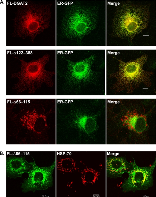
Deletion of the two transmembrane domains disrupts the ER localization of DGAT2. A, COS-7 cells were co-transfected with FL-DGAT2, FL-Δ122–388, or FL-Δ66–115 and the ER marker, ER-GFP (a generous gift from Dr. Erik Snapp, Albert Einstein College of Medicine of Yeshiva University). ER-GFP consists of the bovine prolactin signal sequence fused to the N terminus of GFP and a C-terminal ER retention sequence (KDEL) (30). After fixation and permeabilization, cells were stained with anti-FLAG. B, COS-7 cells expressing FL-Δ66–115 were stained with anti-FLAG and anti-HSP70 to visualize mitochondria. Scale bars, 10 μm.

