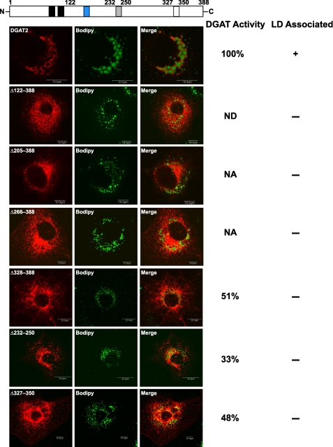
FIGURE 7.
The C terminus is required for the interaction of DGAT2 with lipid droplets. COS-7 cells transiently expressing FL-DGAT2 and FL-DGAT2 mutants with the indicated alterations were treated with 0.5 mm oleate for 12 h and then stained with anti-FLAG and BODIPY 493/503 to visualize lipid droplets. The gray and white boxes represent a potential amphipathic α-helix and proline knot motif, respectively. The blue box is the highly conserved His-Pro-His-Gly sequence (amino acids 161–164) that is part of the active site of DGAT2. Scale bars, 10 μm. In vitro DGAT activities of the DGAT2 mutants were determined and compared with FL-DGAT2 (100% active). DGAT activity was not detectible (ND) for Δ122–388. NA, not assayed.

