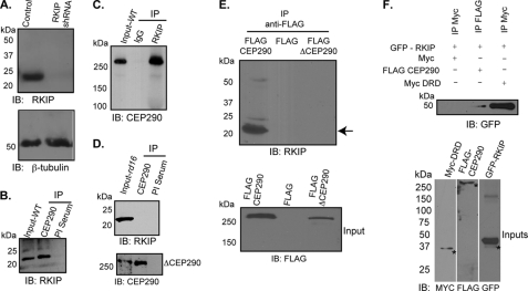
FIGURE 1.
Cep290 interacts with Rkip. A, immunoblot (IB) analysis of protein extracts from control and RKIP-shRNA expressing hTERT-RPE1 cells was performed using anti-Rkip (upper panel) or anti-β-tubulin (lower panel) antibodies. Apparent molecular mass is denoted in kDa. B and C, IP was performed with antibodies to Cep290 (B) or Rkip (C) using mouse retinal extracts. IP with preimmune (PI) serum or normal rabbit IgG was used as negative control. Input represents 10% of the amount of protein used for IP. D, IP using anti-Cep290 antibody (upper panel) from rd16 mouse retina revealed no Rkip-immunoreactive band. Lower panel shows the efficacy of IP using the Cep290 antibody, as ΔCep290 can be immunoprecipitated from rd16 mouse retina. E, COS7 cells were transiently transfected with constructs encoding FLAG epitope, Flag-Cep290, or Flag-ΔCep290. Protein extracts were subjected to IP using anti-FLAG antibody and immunoblotting using anti-Rkip or anti-FLAG antibodies. Arrow indicates the Rkip-immunoreactive band in the FLAG-Cep290 expressing cell extracts. Lower panel shows that FLAG-CEP290 and FLAG-ΔCEP290 proteins were expressed in the cells. F, lysates from COS7 cells transiently transfected with GFP-Rkip, Myc-DRD, and Flag-CEP290 were subjected to IP using the indicated antibodies followed by immunoblotting with anti-GFP antibody. Asterisks represent specific immunoreactive bands. Lower panel shows input samples of the different extracts used for IP.

