FIGURE 2.
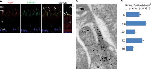
Rkip primarily localizes to cilia. A, retinal sections from WT mice were stained with anti-Cep290 (green) and anti-Rkip (red) antibodies. Merge image shows co-localization of Rkip and Cep290 at the TZ (arrows). Right panel in the merged image shows a magnified image of the co-localization of Rkip and Cep290 at the junction between the inner and outer segments (ONL, outer nuclear layer). B, immunogold labeling of Rkip in mouse photoreceptors reveals broad expression of Rkip in photoreceptors with predominant staining in the apical inner segment (AIS) and TZ. CEN, centriole; BB, basal body. C, histogram shows quantitative analysis of the labeling in the different regions of the photoreceptors. Electron micrographs were analyzed for immunogold particles in a defined square area of the different regions (AIS, BB, and TZ). The number of gold particles counted across various sections was plotted as average number of particles in the region counted. At least 10 cells in each section across 10 different sections were used for analyzing the gold particle density.

