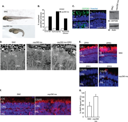
FIGURE 4.
Depletion of cep290 in zebrafish is associated with morphological anomalies and retinal defects. A and B, injection of cep290-MO and not a SNC-MO results in ciliary defects in zebrafish embryos, including curly tail, pericardial effusion, hydrocephaly, and microphthalmia (quantified in B). C, left panel, retinal cryosections from zebrafish embryos injected with cep290-mo or SNC were stained with anti-Cep290 antibody (green). Nuclei are stained with Hoechst (blue). ONL, outer nuclear layer; INL, inner nuclear layer. Right panel shows immunoblot (IB) analysis of lysates of zebrafish injected with SNC or cep290-MO using anti-Cep290 or actin antibody (loading control). D, transmission EM of retinal sections from 4 dpf zebrafish embryos injected with SNC or cep290-MO or co-injected with cep290-MO and mRNA encoding the DRD (cep290-mo+DRD) was performed to detect the effect on photoreceptor development. Arrows indicate loss of photoreceptor OS due to knockdown of cep290. E, effect of loss of cep290 on the expression of photoreceptor-specific proteins ZPR1 and ZPR3 (red) was examined by immunofluorescence analysis of zebrafish embryos treated with SNC or cep290-MO. Nuclei were stained with Hoechst (blue). F, SNC or cep29-MO-treated zebrafish retinas were analyzed for Rkip expression by immunofluorescence using anti-Rkip antibody. Histogram in panel G depicts the up-regulation of Rkip levels following depletion of cep290 as compared with controls. The data are representative of three independent experiments with at least 20 embryos analyzed in each experiment. AFU, arbitrary fluorescent units.

