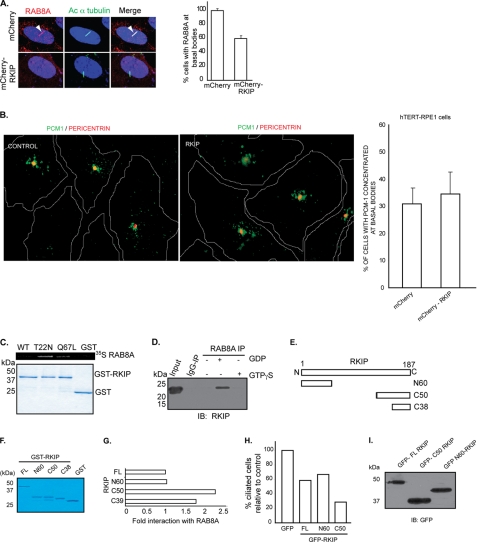
FIGURE 7.
Overexpression of Rkip affects Rab8A localization in cells. A, confocal immunofluorescence of mCherry-Rkip or mCherry alone hTERT-RPE1 stably overexpressing cells stained with anti-Rab8A antibody (represented in red). Rab8A positive staining at cilia (arrow) was assessed by co-localization with acetylated (Ac) α-tubulin (represented in green). There is a considerable decrease in the number of cells with Rab8A at the cilia in Rkip-overexpressing cells (right panel; histogram). Data represent mean ± S.D. (p < 0.005). B, overexpression of Rkip does not seem to affect Pcm-1 distribution in hTERT-RPE1 cells. Localization of Pcm-1 around the basal bodies was assessed by co-localization with Pericentrin (represented as red) in hTERT-RPE1 cells stably expressing mCherry-Rkip (Rkip) or mCherry alone (control). No major difference was observed between the two groups, as shown in the right panel. Data represent mean ± S.D. from more than 150 cells analyzed in each group in three independent experiments. C, GST pulldown assay was performed using GST-Rkip and in vitro translated 35S-Rab8A WT, GDP-locked (T22N), and GTP-locked (Q67L) mutants. Interaction was assessed by autoradiography (upper panel). Lower panel shows Coomassie staining of the proteins used in the assay. D, bovine retinal lysates (∼300 mg), prepared in the presence of GDP or GTP non-hydrolyzable analogs were subjected to IP using Rab8A antibody or IgG (control). Precipitated proteins were analyzed by SDS-PAGE and immunoblotting (IB) using anti-Rkip antibody. E, schematic representation of different deleted variants of Rkip utilized in the GST pulldown assay. F, Coomassie Blue-stained gel of the Rkip variants after purification from E. coli. G, quantitative analysis of the interaction between Rkip (full-length; FL and deleted variants) and 35S-Rab8A-T22N in GST pulldown assay. Data represent mean of three independent experiments. H, quantitative analysis of the number of ciliated hTERT-RPE1 cells after transient overexpression of different GFP-Rkip variants or GFP alone (control). Data represent mean of three independent experiments (n >200). I, lysates from transientely transfected hTERT-RPE1 cells with GFP-tagged variants of Rkip were analyzed by SDS-PAGE and immunoblotting with GFP antibody.

