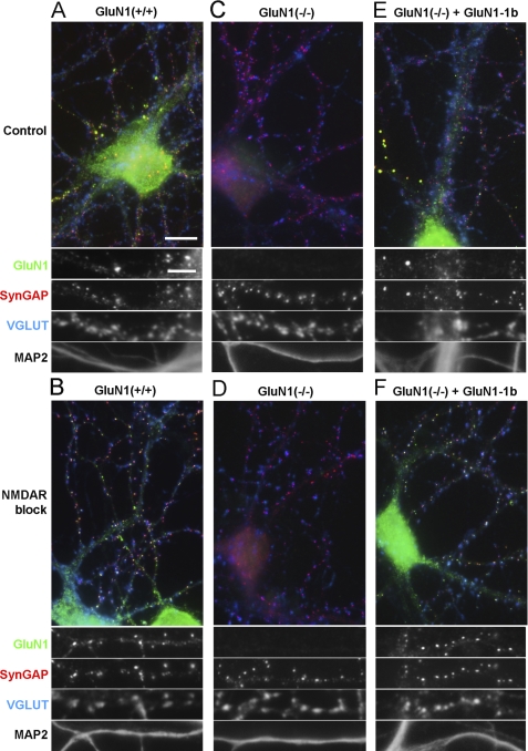
FIGURE 1.
Rescue of GluN1 synaptic clustering in GluN1(−/−) neurons transduced with lentivirus expressing GluN1-1b. Low density hippocampal cultures from wild type (A, B) or GluN1(−/−) (C–F) mice were fixed at 14–15 DIV and immunostained for GluN1, the excitatory postsynaptic component SynGAP, the excitatory presynaptic marker VGLUT, and the dendritic marker MAP2. As indicated, cultured neurons were treated with a mixture of the NMDAR blockers APV (100 μm) and MK801 (5 μm) from 6 DIV, to homeostatically increase synaptic clustering of NMDA receptors (B, D, F). GluN1(−/−) hippocampal neurons exhibited no immunoreactivity for GluN1 (C, D). GluN1(−/−) neurons transduced at 3 DIV with lentivirus expressing GluN1-1b under the control of the synapsin I promoter showed successful rescue of GluN1 immunofluorescence (E, F). No changes in SynGAP, VGLUT or MAP2 immunoreactivity were observed in the GluN1(−/−) transduced neurons compared with wild type neurons. Quadruple staining of hippocampal cultures from GluN1 wild type and GluN1(−/−) mice transduced with GluN1-1b showed in both cases a similar distribution of GluN1. In control conditions, GluN1-1b like wild type GluN1 formed a few bright non-synaptic clusters co-localizing with SynGAP but not VGLUT, shown enlarged in panels A and E, and a greater number of synaptic clusters co-localizing with SynGAP and VGLUT1 (shown enlarged in Fig. 3A for GluN1-1b). Long-term NMDAR blockade increased synaptic accumulation of rescued GluN1-1b (F) like that of wild type GluN1 (B). Scale bars: 10 μm for color overlays, 5 μm for single channel enlarged regions.

