FIGURE 4.
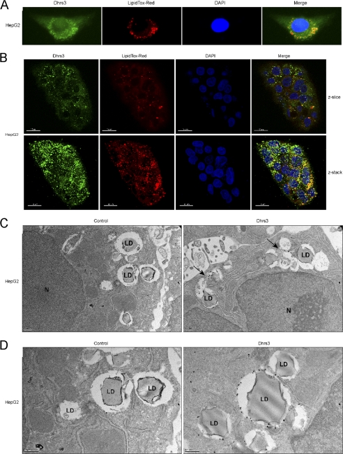
DHRS3 associates with lipid droplets in hepatocytes. A, immunofluorescence staining of endogenous DHRS3 in HepG2 cells. LipidTOX Red marks the lipid droplets, and DAPI marks the nucleus. B, confocal scans of endogenous DHRS3 in HepG2 cells. Both a single z-slice and the composite z-stack are shown. LipidTOX Red marks the lipid droplets, and DAPI marks the nucleus. Scale bar, 15 μm. C and D, transmission electron microscopy of immunogold-labeled endogenous DHRS3 in HepG2 cells. HepG2 monolayers were fixed, probed with secondary antibody alone (left) or α-DHRS3 primary antibody (right), and visualized by transmission electron microscopy. Representative lipid droplets (LD) and nucleus (N) are indicated. Arrows denote point of interest.

