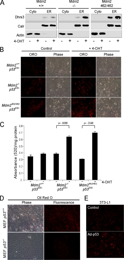
FIGURE 8.
p53 induction of DHRS3 correlates to greater lipid droplet levels. A, mouse embryonic fibroblasts harboring a single p53ER fusion allele and a single p53 null allele (p53 ER/- MEF) cell lines with the indicated Mdm2 genotypes were treated for 24 h with 4-OHT. Cells were harvested and partitioned into cytosolic-enriched (Cyto) or ER-enriched fractions and analyzed by Western blot. Actin is a cytosolic marker, and calreticulin (Calr) is an ER marker. B, p53 ER/- MEF cell lines with the indicated Mdm2 genotypes were treated for 24 h with 4-OHT and stained with Oil Red O (ORO). Cells were visualized for Oil Red O uptake by fluorescence microscopy. Phase contrast image (Phase) is shown for cell density control. C, Oil Red O was extracted from samples in B and analyzed by spectrophotometry. The ratio of total Oil Red O absorbance per milligram of protein was used to quantitate total values. p values are shown for statistically significant samples. Error bars represent S.E. D, p53 +/+ and −/− MEF cells were grown to confluence and stained with Oil Red O. Stain uptake was visualized by phase-contrast and fluorescence microscopy. E, 3T3-L1 preadipocytes were either transduced with adenovirus expressing p53 or left untreated. Cells were stained with Oil Red O 24 h post-transduction.

