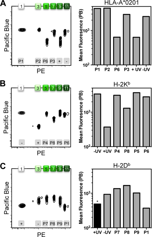
FIGURE 2.
Detection of major histocompatibility complexes on fluorescent beads. Soluble MHC molecules were ligand-exchanged with peptides P1 (GILGFVFTL), P2 (NLVPMVATV), P3 (FLPSDFFPSI), and P6 (SIINFEKL) in combination with the human HLA-A*02:01 (A); peptides P4 (SVLAFRRL), P5 (SIYRYYGL), P6 (SIINFEKL), and P8 (ASNENMETM) in combination with the murine MHC product H-2Kb (B); or peptides P1 (GILGFVFTL), P7 (SAVSNLFYV), P8 (ASNENMETM), and P9 (ASFVNPIYL) in combination with the murine MHC molecule H-2Db (C). As controls, the different MHCs were treated with (+) or without (−) UV irradiation in the absence of replacement peptide. The resulting MHC products were captured on streptavidin-coated Pink beads that are increasingly fluorescent in a distinct series of peaks. For the six different conditions tested per MHC, the use of only the odd-numbered bead populations (i.e. 1, 3, 5 7, 9, and 11) was sufficient. The beads were probed for intact MHC with anti-β2m conjugated with PB. The black bar (*) indicates that the H-2Db control reaction was incubated at 50 °C to drive complex disintegration to completion. Bead populations are numbered on the top x axis, whereas the employed exchange conditions are indicated on the bottom x axis.

