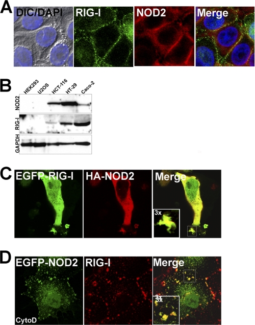
FIGURE 1.
RIG-I and NOD2 colocalize at cell-cell junctions and membrane ruffles. A, HT-29 cells grown under confluent conditions were fixed and stained with anti-RIG-I (green) and anti-NOD2 (red) antibodies. At left are shown differential interference contrast (DIC) and DAPI (nuclei, blue) merged images. B, lysates (50 μg) from the indicated cell types were immunoblotted for RIG-I, NOD2, and GAPDH as a loading control using a LI-COR Odyssey infrared imaging system. C, HEK293 cells were transfected with EGFP-RIG-I and HA-NOD2 and fixed and stained for HA (red). D, Caco-2 cells were transfected with EGFP-NOD2 and 48 h post-transfection, exposed to 10 μm cytochalasin D (cytoD) for 60 min. Cells were then fixed and stained for endogenous RIG-I (red). Data are representative of experiments performed at least three times.

