FIGURE 7.
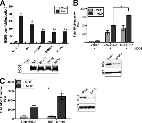
RIG-I suppresses NOD2 in intestinal cells. A, luciferase assay from HEK293 cells transfected with the indicated HA-NOD2 constructs and ISG56-Luciferase reporter. Forty-eight hours post-transfection, cells were stimulated with Sendai virus (100 HAU/ml) for 16 h and luciferase activities measured. Results are expressed as fold induction of ISG56-luciferase relative to that of mock-infected cells after normalizing to Renilla luciferase. Immunoblots of HA-NOD2 expression are shown in the bottom panel. B, top, luciferase assay (expressed as fold NF-κB induction versus vector controls) from HeLa cells stably expressing control or RIG-I shRNA and transfected with HA-NOD2 (250 ng) plus NF-κB promoter luciferase (100 ng) and MDP (10 μg/ml) for 24 h. Bottom, immunoblots from lysates shown at top. C, left, luciferase assay (expressed as fold NF-κB induction versus untransfected controls) from HT29 cells transfected with control (Con) or RIG-I siRNAs plus NF-κB promoter luciferase (250 ng). Forty-eight hours following transfection, cells were exposed to MDP (20 μg/ml) for 14 h and luciferase activity measured. Right, immunoblots from lysates at left. Data are shown as mean ± S.D. Asterisks indicate p values < 0.05 (*) or <0.001 (**).

