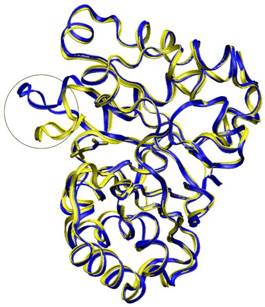
Figure 1.
Superimposed apo- (in yellow) and holo- (in blue) crystal structures of triosephosphate isomerase. PDB code 1YPI and 2YPI, respectively [116]. The 11 residue-loop composed of binding site is the only region that has large motion upon ligand binding (in circle).

