Figure 1.

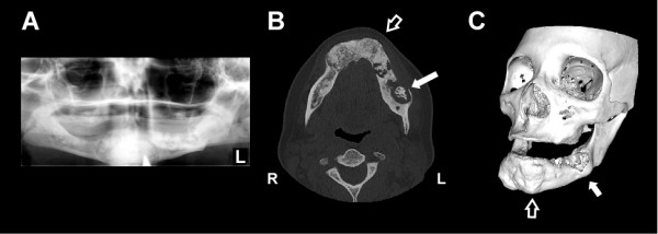
Radiographic evidence of Actinomyces osteomyelitis complicating florid cemento-osseous dysplasia (FCOD). (A) Panoramic radiograph demonstrating mixed radiolucent and radiopaque lesions in the mandible with "cotton wool" appearance. Lesions are well demarcated with a radiolucent ring in all four quadrants though they are more subtle in the maxilla (B) Axial CT scan image showing hypertrophic, sclerotic and heterogeneous changes of FCOD within the mandible (open arrow). There is a large lytic lesion in the body of the left mandible with loss of bone at its lateral aspect and central sclerosis consistent with infection (solid arrow). (C) 3-dimensional CT image of generalized bony changes with expansion to maxilla and mandible consistent with FCOD (open arrow, corresponding to same location in panel A). There is focal erosion of left mandible in area of Actinomyces infection (solid arrow). CT images were reformatted with OsiriX imaging software (OsiriX Foundation).
