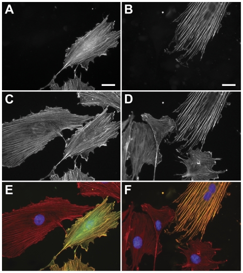
Figure 1. Morphology of pEGFP-Lifeact and pEYFP-actin transfected rat embryonic fibroblast cells (REF52wt).
Lifeact (A) and Actin (B) co-localize with Alexa647-phallodin staining (C,D). Phalloidin staining and EGFP-Lifeact expressing cells are corresponding well in their actin morphology. Cells expressing the EYFP-actin fusion protein reveal more pronounced stress fibers compared to the surrounding non-transfected cells. (E,F) Merged images. (Scale bar: 25 µm).

