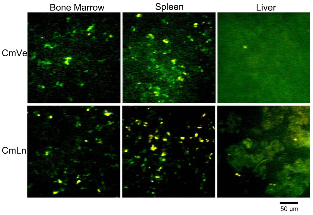
Figure 3.
Confocal scanning images of bone marrow, spleen and liver tissues of rats after 6 h of intravenous administration of curcumin vesicles (CmVe) and curcumin lipid nanospheres (CmLn). Yellow flourescence shows the presence of curcumin in the tissues (From Sou et al., reproduced with permission) (74).

