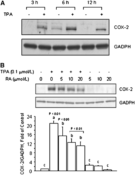
FIGURE 4 .
RA reduces TPA-induced COX-2 expression in nonmalignant breast epithelial MCF10A cells. (A) Time course of COX-2 protein induction by TPA. MCF10A cells were cultured in basal medium plus vehicle (−) or in the presence (+) of TPA (0.1 μmol/L) for 3, 6, and 12 h. (B) Effects of RA on basal and TPA-induced COX-2 protein. MCF10A cells were pretreated for 1 h with RA and then cultured for 6 h in the presence of TPA (0.1 μmol/L), RA (5, 10, 20 μmol/L), or their combination. (A,B) Bands represent COX-2 and GADPH (loading control) immunocomplexes. Values are means + SE of triplicates from 2, n = 2, independent experiments. Means without a common letter differ, P < 0.05.

