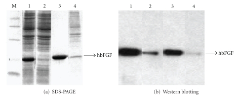
Figure 3.
(a) 12% SDS-PAGE analysis of fractions from purification of hbFGF. M: Molecular weight marker; lane 1: cell lysate in 8 M urea, PBS pH 7; lane 2: flow through; lane 3: peak 1; lane 4: peak 2. (B) Western blotting of cell lysate and fractions from purification of hbFGF. Lane 1: cell lysate; lane 2: flow through; lane 3: peak 1; lane 4: peak 2.

