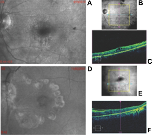
Figure 1.
(A) Representative F-10 blue laser imaging of one eye with pre-retinal wrinkling; (B) Infra-red fundus photograph of the same patient showing the position of the scan in image (C); (C) optical coherence tomography scan of the same eye, which shows pre-retinal wrinkling. (D) Representative F-10 blue laser imaging on one eye with macular pucker and macular hole; (E) Infra-red fundus photograph of the same patient showing the position of the scan in image (F); (F) optical coherence tomography of the same eye which shows macular pucker.

