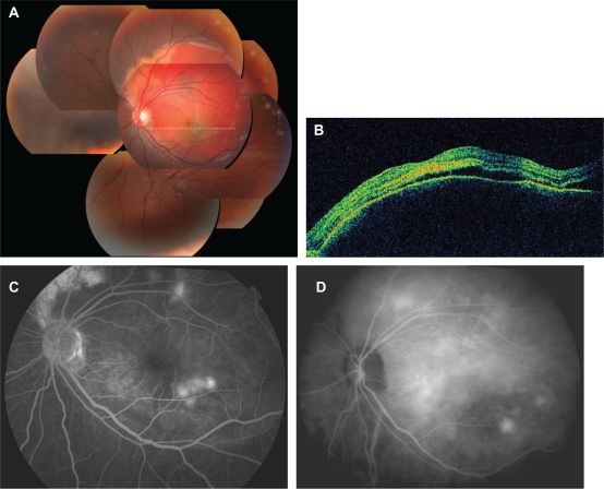
Figure 1.
Patient with a choroidal osteoma and choroidal neovascularization before treatment. (A) Fundus photograph showing 5-disc-diameter hemorrhage under the retinal pigment epithelium. (B) Horizontal optical coherence tomographic image, showing pigment epithelial detachment and serous retinal detachment. (C, D) Angiography: fluorescein angiography (C) and indocyanine angiography (D) show several points of dye leakage around the fovea.

