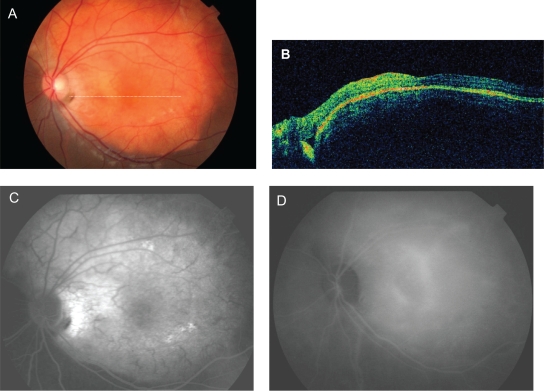
Figure 3.
Patient with a choroidal osteoma and choroidal neovascularization 18 months after two intravitreal injections of bevacizumab. Fundus photograph (A), horizontal optical coherence tomographic scan (B), fluorescein angiography indocyanine angiography (C), and indocyanine angiography (D) all show absence of subretinal fluid and leakage points.

