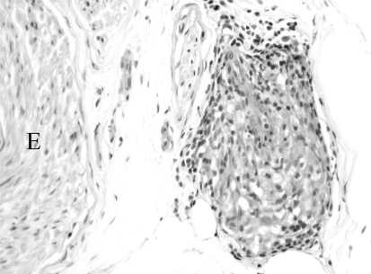
Fig. (1).
A hematoxylin- and eosin-stained paraffin section of a superficial peroneal sensory nerve biopsy specimen reveals a granuloma consisting of epithelioid histiocytes surrounded by a rim of lymphocytes. Giant cells are not seen and may not be readily apparent in most nerve granulomas. The granuloma is in the epineurium adjacent to the endoneurium (E).

