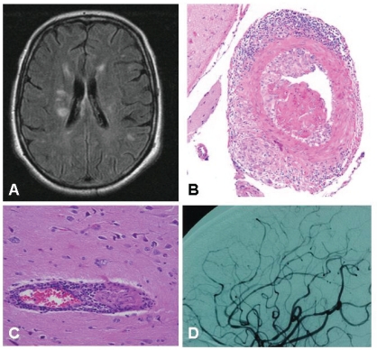
Fig. (1A).
Multiple, non-specific, T2 hyperintense lesions in a 63- year old patient with suspected primary angiitis of the CNS who presented with headache and cognitive impairment. B) Granulomatous pattern of primary angiitis of the central nervous system. Transmural inflammation involves a muscular artery of the leptomeninges with prominent mononuclear (upper) and granulomatous (lower) adventitial inflammation as well as intimal injury with focal fibrin thrombus formation (hematoxylin and eosin 20×). Courtesy of Dr Carlo Salvarani. C) Inflammatory involvement of a small vessel. Courtesy of Dr Leonard H Calabrese. D) Multiple areas of irregular stenosis and ectasia in a 44year-old patient with biopsyproven PACNS. Courtesy of Dr Leonard H Calabrese.

