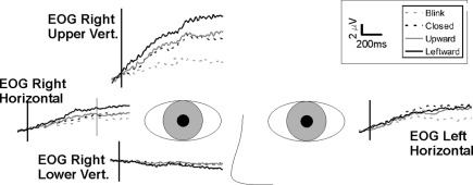
Figure A1.
Average activity in electro-oculogram (EOG) electrodes. Grand averages (n = 14) for each of the four EOG electrodes, are shown relative to eye position. Note that there is persisting positive-going activity in the upper vertical electrode and no (negative-going) activity in the lower EOG electrode. The activity shows a similar time course, but smaller amplitude, in the lateral EOG electrodes. When slow activity is observed in frontal electrodes, there is always the concern that this may have resulted from eye movement or drift. In three of the four EOG electrodes there was a large positivity, with a gradient of activity. It was largest in the upper vertical, and was present albeit to a lesser extent in the bilateral horizontal eye channels. The time course of the positivity was identical to that observed in the frontal EEG channels. Importantly however, the lower vertical EOG channel, located below the right eye, did not show a reflected negative activity as it would for any vertical eye drifts, suggesting that observed activity on the other EOG (and frontal EEG) electrodes represented EEG activity.

