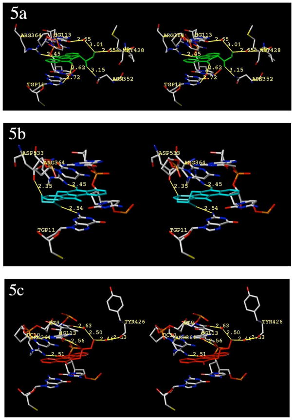
Figure 5.
Hypothetical models of the ternary complexes with Top1, DNA, and diols 12a and 12b. All views are from the major groove, scissile-strand side. The normal binding mode of compound 12a (green ligand) is shown in 5a. The flipped binding mode of compound 12a (cyan ligand) is shown in 5b. The normal (only) binding mode of compound 12b (red ligand) is shown in Figure 5c. All other structures are colored by element, water molecules are shown as red spheres and relevant substructures are labeled. Hydrogen bonds and polar contacts are shown and labeled. Distances (in Å) are from heavy atom to heavy atom. The diagrams are programmed for wall-eyed (relaxed) viewing.

