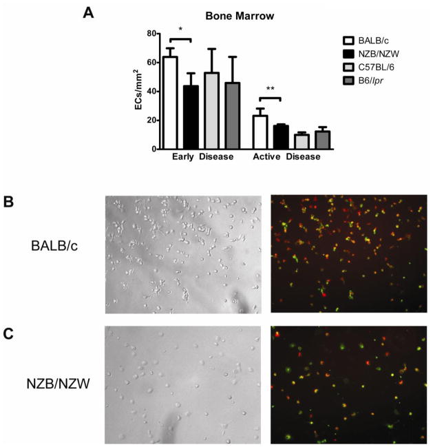
Figure 3. NZB/W EPCs exhibit impaired capacity to differentiate into mature ECs.
Bone marrow-derived EPCs were cultured under proangiogenic conditions and incubated at different time-points during culture with Dil-ac-LDL and BS-1-FITC. Mature endothelial cells were identified by co-staining of BS-1 and ac-LDL. (A) Bar graphs represent the number of mature endothelial cells per centimeter at day 7, when comparing NZB/W and B6/lpr bone marrow-derived EPCs with control EPCs, at early and active disease time-points. Results are mean ± SEM of 3 or 4 independent experiments; *p<0.05, **p<0.01. (B), (C) Results are representative images obtained from BALB/c (B) and NZB/W (C) EPCs cultured under proangiogenic conditions for 7 days. Left panels show bright field images and right panels show images obtained by fluorescent microscopy. NZB/W EPCs show decreased ability to differentiate into endothelial cells, when compared to BALB/c mice. DiI-Ac-LDL is red while BS-1 lectin is green. Total magnification is ×100.

