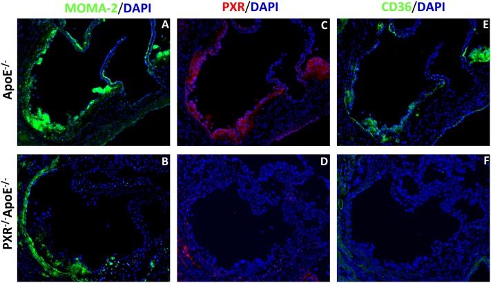
Fig.5.
Immunohistochemistry of PXR and CD36 in atherosclerotic lesions of apoE−/− and PXR−/−apoE−/− mice. Sections of atherosclerotic lesion area in the aortic root of apoE−/− and PXR−/−apoE−/− mice were stained with anti-monocytes/macrophages (MOMA-2) (A, B), anti-PXR (C, D), or anti-CD36 (E, F) primary antibodies, followed by fluorescein-labeled secondary antibodies. The nuclei were stained with DAPI (blue). Magnification, ×100.

