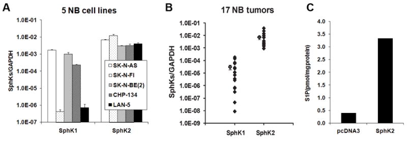
FIGURE 1.

SphK2 was abundantly expressed in NB cells and tissues. A and B, The mRNA expression of SphK1 and SphK2 in 5 NB cell lines (A) and 17 human NB tissues (B) by quantitative real-time PCR. The mean values for SphK1 and SphK2 in B were shown at the left side of each column. C, The S1P synthesis in SK-N-AS cells transfected with pcDNA3 or SphK2 plasmid. Representative data from two independent experiments was shown.
