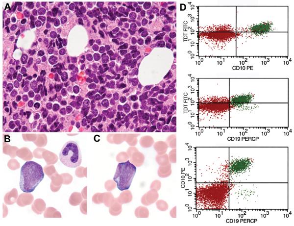
Figure 2.
B-Lymphoblastic Leukemia with IGH-BCL2 and MYC Rearrangements
The bone marrow core biopsy from case 20 showed medium-sized cells with round nuclei and finely dispersed chromatin in a background of extensive cellular necrosis (A). The peripheral blood contained a significant population of blasts with round to irregular nuclei, prominent nucleoli, dispersed chromatin and deeply basophilic cytoplasm (B-C). Immunophenotyping of the circulating leukemic cells by flow cytometry showed a population of CD19+, CD10+, terminal deoxynucleotidyl transferase (TdT)+ B lymphoblasts (D) that were CD45dim+ and negative for CD20 and surface light chain (not shown).

