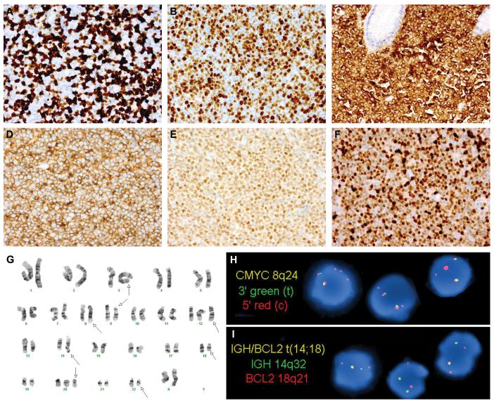
Figure 4.
Immunophenotypic, Cytogenetic and Molecular Genetic Features of Double-Hit Lymphoma
Two examples of double-hit lymphoma (DHL) demonstrating a Ki-67 proliferation index (PI) of <95%: a retroperitoneal lymph node biopsy from case 5, classified as B-cell lymphoma, unclassifiable, with features intermediate between diffuse large B-cell lymphoma and Burkitt lymphoma (BCLU), with an overall Ki-67 PI of 60% (A); and an abdominal lymph node biopsy from case 14, classified as diffuse large B-cell lymphoma, not otherwise specified (DBLCL-NOS), with an overall Ki-67 PI of 80% (B). The vast majority of DHL cases expressed Bcl2 by immunohistochemistry for the commonly used antibody, clone 124 (C: stomach polyp, case 1). All DHL cases were of germinal center origin, expressing CD10, Bcl6, or both, as in this cervical lymph node biopsy from case 12 (D: CD10, E: Bcl6). Mum1 was expressed in 8/19 DHL cases, including this axillary lymph node biopsy from case 2 (F). Bone marrow cytogenetic analysis from case 4 showed a complex karyotype (G, arrows), as did all DHL cases. In addition to the t(8;22) and t(14;18), this case contained additional unknown material in the 3q2?7 region (G, arrow indicating chromosome 3). Subsequent fluorescence in situ hybridization (FISH) analysis confirmed a rearrangement involving BCL6 at 3q27 (not shown). Interphase FISH analysis of paraffin-embedded stomach polyp tissue from case 1 confirmed both a MYC rearrangement (H: dual-color, split-apart probe) and an IGH-BCL2 fusion (I: dual-color, dual fusion probe). The FISH patterns with both probes were consistent with unbalanced rearrangements (H-I). This patient’s prior low-grade follicular lymphoma lacked both MYC and BCL2 rearrangements (not shown), suggesting that both translocations were acquired at the time of DHL transformation.

