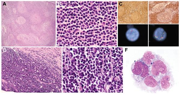
Figure 6.
Pre-Existing Low-Grade Follicular Lymphomas with Unusual Features
Examination of prior low-grade follicular lymphomas in 2 patients revealed findings unusual for this diagnosis. In case 10 with testicular double-hit lymphoma (DHL; illustrated in Fig. 3D), review of a supraclavicular lymph node biopsy from 2 years earlier showed architectural effacement by a follicular proliferation of B cells (A). Cells within and outside of follicles were small with blastoid features, including round nuclei, evenly dispersed chromatin and absent nucleoli (B); typical centrocytes were not identified. Both follicular and extrafollicular B cells were strongly CD20+, CD10+ and Bcl2+ (C, upper left). The follicular component weakly co-expressed Bcl6, but showed absent follicular dendritic cell staining (not shown). Ki-67 stain showed an unusually high proliferation index (PI) of approximately 40% within follicles and 90% outside of follicles (C, upper right). Fluorescence in situ hybridization (FISH) analysis with dual color break apart probes revealed a MYC rearrangement (C, lower left) and 8 copies of BCL2 (C, lower right), the same genetic abnormalities seen in the subsequent DHL. In case 11, core needle biopsies of a retroperitoneal mass showed a CD10+, Bcl6+, Bcl2+ B-cell neoplasm with a diffuse growth pattern (absent staining for follicular dendritic cell antigens), a focal starry-sky pattern (D) and patchy necrosis. The cells were small to medium-sized; some were centrocyte-like, but the majority had oval nuclei with finely dispersed chromatin and absent nucleoli (E), prompting a diagnosis of diffuse grade 1-2 FL with blastoid features. Tissue from the initial biopsies was not available for cytogenetics or FISH analysis. Six months later, the patient relapsed with a CD10+ B-cell lymphoma involving the central nervous system (F), confirmed by FISH to have an IGH-BCL2 fusion as well as a MYC rearrangement (FISH analysis not shown).

