
Figure 4.
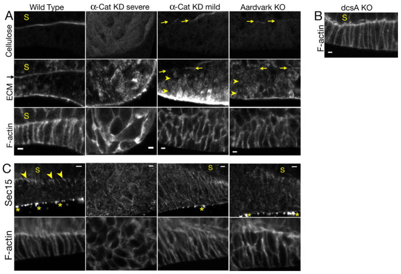
A) Confocal sections of the tip epithelium in culminants of the indicated cells. Arrows indicate deposition of small amounts of extracellular cellulose and EcmA/B in a nascent stalk tube. Arrowheads indicate intracellular accumulation of EcmA/B.
B) Confocal section of the tip epithelium in a culminant of cellulose synthase (dcsA) knockout cells (12).
C) Confocal sections of tip epithelia in culminants of the indicated cells. Arrowheads indicate Sec15 localization. Asterisks indicate non-specific signal on the exterior of the culminant (Fig. S8).
Scale bars: 2 μm.