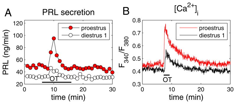
Figure 4.
Comparison of the oxytocin-induced responses in perifused lactotrophs obtained from female rats on the morning of diestrus 1 (open symbols) and the afternoon of proestrus (closed red symbols). (A) Oxytocin application (100 nM for 10 min) evoked a larger prolactin-releasing effect from proestrus cells. Samples were collected every minute. (B) Oxytocin application (100 nM for 2 min) elicited a larger increase in the free intracellular Ca2+ concentration, as measured by the fura-2 fluorescence ratio. See (50) for details and a full description of experimental procedures.

