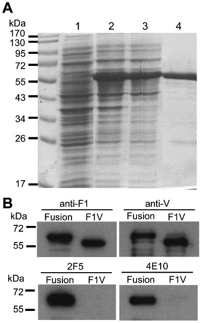
Fig. 1.

Analysis of F1-V-MPR649-684 expressed in E. coli. (A) SDS-PAGE analysis of F1-V-MPR649-684 expression in E. coli. Lane 1, pre-induction control E. coli proteins; lane 2: total cell proteins 2 h post-induction; lane 3: soluble cell proteins after disruption of cells; and lane 4: F1-V-MPR649-684 purified by metal affinity chromatography. (B) Immunoblot analysis of E. coli-expressed F1-V and F1-V-MPR649-684. “Fusion” represents F1-V-MPR649-684. Polyclonal rabbit Abs to F1 (upper left) or V (upper right), or gp41-specific human mAbs 2F5 (bottom left) or 4E10 (bottom right) were used for detection. F1-V-MPR649-684 was detected by all Abs, whereas F1-V was recognized by anti-F1 and -V Abs only.
