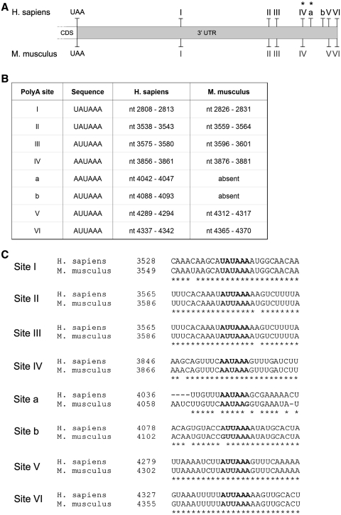
Figure 2.
Structure of the FMR1 3′-UTR in human and mouse brain. (A) Schematic representation of the putative poly(A) sites, taking into account both canonical and not-canonical sites (48) in human and mouse. Asterisks mark canonical poly(A) sites. (B) Nucleotide position and consensus sequence of the poly(A) sites in both human and mouse. (C) Alignment between the regions containing the putative poly(A) signals in human (accession number NM_002024) and mouse (accession number NM_008031). Consensus sequences are bolded. The alignment was performed using the ClustalW software.

